Vaping-Associated Lipoid Pneumonia: Case Report of 29-Year-Old Male with Right Middle Lobe Pneumonia at IRC Islamabad
Islamabad Respiratory Clinic (IRC), G-11 Markaz, Islamabad | Published: 11 Oct 2026 | Updated: 11 Oct 2026 | Patient consent obtained, de-identified for education
Written and Reviewed by: Dr. Muhammad Israr — Head of Pulmonology, Consultant Respiratory Physician & Interventional Pulmonologist, Islamabad Dr. Jehangir Khan — Consultant Pulmonologist & Interventional Pulmonologist, Islamabad Consultant Pulmonologists in Islamabad | IRC G-11 Markaz
1. Introduction
Lipoid pneumonia is an uncommon inflammatory lung condition where lipids accumulate in alveoli. Exogenous lipoid pneumonia has been reported after inhalation of oil-based substances, including mineral oils and components of vape liquids. Vaping-associated lung injury (EVALI) emerged as a clinical entity with presentations ranging from ground glass opacities to consolidations.
We present a 29-year-old male vape user evaluated at Islamabad Respiratory Clinic, G-11 Markaz, Islamabad, with cough, chest pain and shortness of breath, with imaging showing right middle lobe pneumonia and possible lipoid pneumonia, with subsequent clinical improvement.
2. Case Presentation
A 29-year-old male, known vape user, presented to IRC Islamabad with:
Cough for several days
Chest pain, localized
Shortness of breath on exertion
No high-grade fever reported at presentation, no hemoptysis
Past history: regular vaping use, no known asthma, no tuberculosis contact
On examination, respiratory system examination revealed reduced breath sounds and crackles in right lower and middle zones. Systemic examination was unremarkable. Oxygen saturation was monitored and routine labs were obtained.
3. Initial Investigations
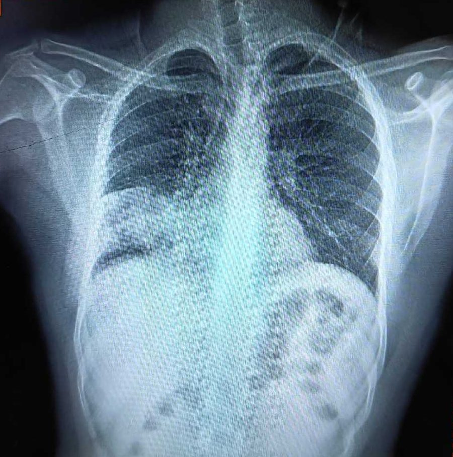
Chest X-ray: pulmonary consolidation consistent with pneumonia (see Figure 1)
Complete blood count, CRP, infection workup as per clinic protocol
Sputum examination and cultures where indicated to exclude bacterial infection and tuberculosis
Spirometry planned after acute phase
4. Chest X-Ray and HRCT Chest Findings
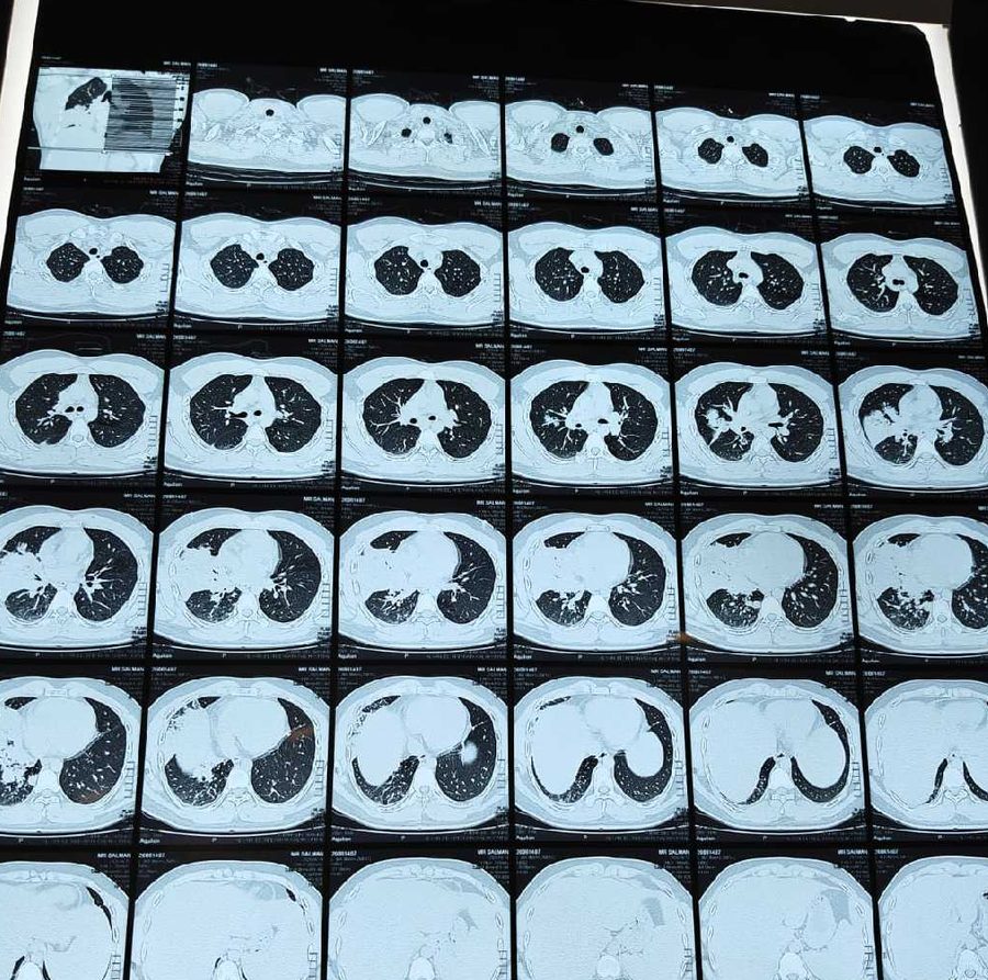
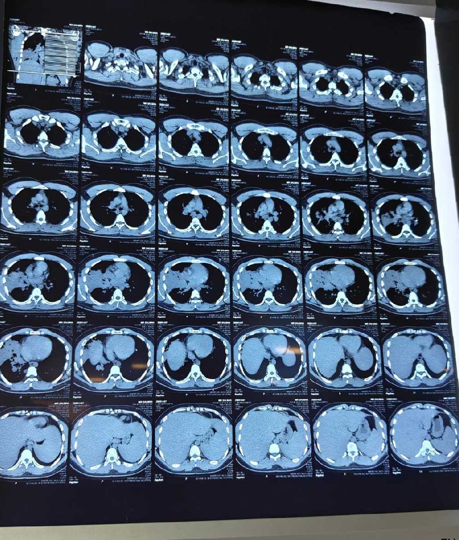
A 29-year-old male vape user presented to Islamabad Respiratory Clinic (IRC), G-11 Markaz, Islamabad, with cough, chest pain and shortness of breath. Initial chest radiography showed pulmonary consolidation consistent with pneumonia. HRCT chest was subsequently performed to characterize the pulmonary abnormality and investigate possible vaping-associated lung injury.
Chest X-Ray at Presentation
Figure 1. Chest radiograph obtained during the initial presentation, demonstrating pulmonary opacity in right middle and lower zones. Clinical correlation required.
HRCT Chest: Lung Window
Figure 2. HRCT chest lung window. Patchy consolidations and ground glass opacities predominantly in right middle lobe and bilateral lower lobes, right greater than left. Findings to be interpreted with full radiology report.
HRCT Chest: Mediastinal Window and Additional Images
Figure 3. HRCT mediastinal window and additional images reviewed as part of clinical assessment. Reported findings raised suspicion of right middle lobe pneumonia with possible lipoid pneumonia. Differential diagnosis including infection required.
Note: Foamy lipid-laden macrophages are not specific for lipoid pneumonia, and their presence alone does not establish the diagnosis. Correlation with complete radiology report, exposure history and evaluation for alternative causes including infection is essential.
5. Bronchoscopy and Laboratory Findings
Bronchoscopy was performed as per indication to evaluate persistent consolidation and to obtain bronchoalveolar lavage (BAL) for analysis.
Bronchoscopy: airway examination noted and BAL obtained from affected lobe
BAL cytology: foamy lipid-laden macrophages observed, which can be seen in lipid-associated inflammatory processes
Microbiology: bacterial cultures, fungal and tuberculosis workup performed to exclude alternative causes
No malignancy seen on cytology
Foamy macrophages can be observed in various conditions and are not diagnostic alone. Diagnosis of lipoid pneumonia requires clinical, radiologic and pathologic correlation and exclusion of infection.
6. Diagnosis
Provisional assessment: Right middle lobe pneumonia with possible vaping-associated lung injury and possible lipoid pneumonia in a 29-year-old vape user. Differential includes community-acquired pneumonia, aspiration pneumonia, organizing pneumonia and EVALI. Final diagnosis was made after clinical correlation and exclusion of alternative causes.
7. Management and Clinical Outcome
Management included:
Counseling for complete vaping cessation and avoidance of oily inhalants
Supportive care, hydration and symptomatic treatment
Antibiotic therapy as per pneumonia protocol while awaiting cultures
Monitoring of oxygenation and clinical parameters
Follow-up imaging and clinic review
Clinical Outcome
At one-month follow-up, the patient was clinically well. Chest examination was clear, and follow-up chest X-ray was reported as normal. This documented clinical and radiological improvement was reassuring. Long-term follow-up and smoking and vaping cessation counseling were advised.
8. Discussion: Lipoid Pneumonia and Vaping
Exogenous lipoid pneumonia results from aspiration or inhalation of lipid-containing substances. Vape liquids contain propylene glycol, vegetable glycerin, flavorings and other additives that may include lipids. When aerosolized and inhaled repeatedly, these can cause inflammatory response in distal airways and alveoli.
HRCT findings in lipoid pneumonia typically include:
Low attenuation consolidations (fat density -30 to -150 HU may be seen but not always)
Ground glass opacities
Predilection for dependent lobes, right middle lobe and lower lobes
Crazy-paving pattern in some cases
EVALI and lipoid pneumonia overlap clinically. Centers for Disease Control and Prevention (CDC) has reported cases of vaping-associated lung injury requiring hospitalization. Vaping cessation is cornerstone of management.
9. Prevention and When to Seek Care in Islamabad
Avoid vaping and e-cigarettes, especially oil-based or unregulated products
Seek pulmonology evaluation if you develop cough, chest pain, shortness of breath within weeks of vaping
Do not self-medicate with steroids or antibiotics without medical evaluation
At IRC Islamabad, evaluation includes detailed history, chest X-ray, HRCT chest, spirometry and bronchoscopy where indicated
10. Frequently Asked Questions
What is lipoid pneumonia?
Lipoid pneumonia is inflammation due to accumulation of lipid material in alveoli. It can be exogenous from inhaled oils or endogenous.
Can vaping cause lipoid pneumonia?
Cases have been reported where vaping was associated with lipoid pneumonia pattern. Risk appears related to constituents of vape liquid and frequency of use. Not all vape users develop lung injury.
How is it diagnosed?
Diagnosis needs combination of exposure history, imaging (HRCT chest), bronchoscopy with lavage showing lipid-laden macrophages, and exclusion of infection and other causes.
Is it reversible?
Many patients show clinical and radiological improvement after cessation of exposure and supportive care, as seen in this case with normal follow-up X-ray at one month. Long-term follow-up is still advised.
Disclaimer: This case report is for medical education and does not replace individual medical evaluation. Images shared with patient consent, de-identified, no personal identifiers included. No identifiable information is present. For persistent respiratory symptoms, consult a qualified pulmonologist at Islamabad Respiratory Clinic, G-11 Markaz, Islamabad or nearest emergency service. If you have severe shortness of breath, chest pain, low oxygen saturation or high fever, seek emergency care immediately.